Contents
Overview
Ultrasound diagnostics, also known as medical ultrasonography or sonography, is a non-invasive imaging technique that utilizes high-frequency sound waves to create real-time visual representations of internal body structures. These sound waves, far above the range of human hearing, are emitted by a transducer and reflect off different tissues and organs within the body. The returning echoes are then processed by an ultrasound machine to generate images, or sonograms, revealing details about organ size, shape, texture, and blood flow. This technology offers a safe and versatile tool for diagnosing a vast array of conditions across specialties like obstetrics, cardiology, radiology, and emergency medicine. Its ability to provide immediate, portable, and radiation-free imaging makes it indispensable in clinical settings worldwide.
🎵 Origins & History
The genesis of ultrasound diagnostics traces back to the early 20th century, building upon discoveries in sonar technology developed for World War I and World War II to detect submarines. Early pioneers like Karl Z. Susman and Douglas Howry began experimenting with reflected ultrasound for medical purposes in the late 1940s and early 1950s, initially focusing on detecting gallstones and brain tumors. The subsequent decades saw rapid advancements in transducer technology, image processing, and Doppler capabilities, transforming ultrasound from a niche tool into a ubiquitous diagnostic modality.
⚙️ How It Works
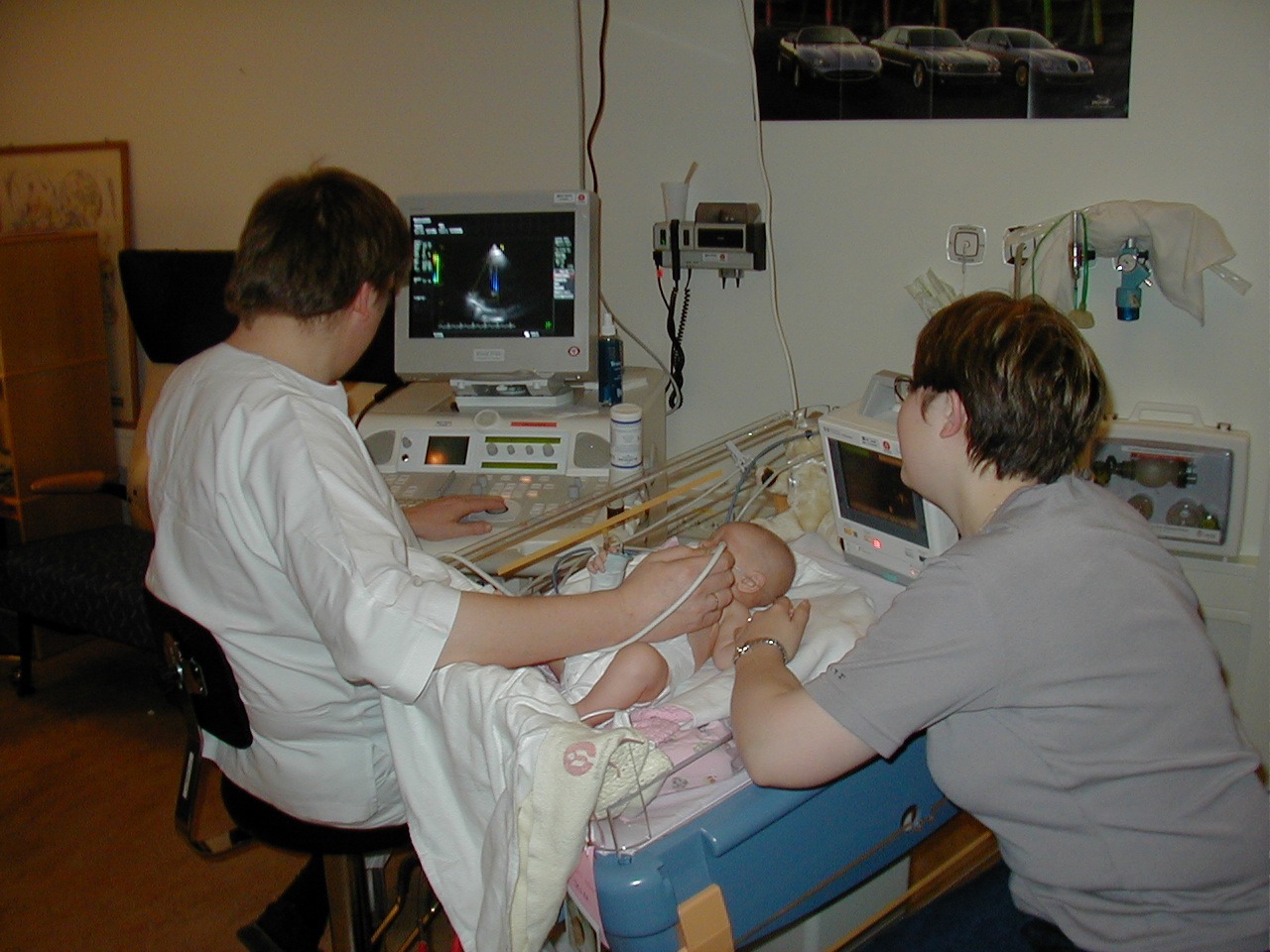
Ultrasound diagnostics operate on the principle of acoustics and echo-location. A handheld device called a transducer emits short pulses of high-frequency sound waves (typically 2-18 MHz) into the body. These sound waves travel through tissues and reflect differently based on the acoustic impedance of the structures they encounter. The transducer then receives these returning echoes, which are converted into electrical signals. These signals are processed by the ultrasound machine's computer, which uses complex algorithms to reconstruct a real-time image on a display screen. Different imaging modes, such as B-mode (brightness mode for anatomical imaging), M-mode (motion mode for tracking movement, especially of the heart), and Doppler ultrasound (for visualizing blood flow and velocity), provide diverse diagnostic information.
📊 Key Facts & Numbers
Key figures in the development of ultrasound diagnostics include Douglas Howry, often called the 'father of diagnostic ultrasound' for his pioneering work in the 1950s with the 'A-scan' and 'B-scan' devices. Ian Donald, a Scottish obstetrician and gynecologist, is credited with developing the first practical diagnostic ultrasound scanner for clinical use in obstetrics in the early 1960s. Joseph Holmes, a radiologist, was instrumental in advancing ultrasound technology for fetal imaging and measurement. Major organizations driving innovation and standardization include the American Institute of Ultrasound in Medicine (AIUM), the Society of Diagnostic Medical Sonography (SDMS), and the World Federation for Ultrasound in Medicine and Biology (WFUMB). Companies like GE Healthcare, Philips Healthcare, and Siemens Healthineers are leading manufacturers of ultrasound equipment.
👥 Key People & Organizations
Ultrasound diagnostics have profoundly reshaped patient care and medical education. Its non-invasive nature and ability to provide immediate results have made it a preferred imaging modality in emergency departments, reducing reliance on more time-consuming or invasive procedures. The widespread availability of ultrasound training programs, offered by institutions like Johns Hopkins University and Stanford University, has democratized its use. Furthermore, the visual nature of sonograms has influenced how medical information is communicated, appearing in documentaries, news reports, and even popular culture, demystifying internal anatomy for the public.
🌍 Cultural Impact & Influence
The current landscape of ultrasound diagnostics is characterized by rapid technological integration and expanding applications. Advances in artificial intelligence are being incorporated to automate image analysis, improve image quality, and assist in diagnosis, with companies like Butterfly Network leading the charge with AI-powered handheld devices. Micro-ultrasound and high-frequency ultrasound are enabling visualization of smaller structures with unprecedented detail, particularly in areas like dermatology and neurology. The development of contrast-enhanced ultrasound (CEUS) is also gaining traction, allowing for better characterization of lesions and improved assessment of blood flow in organs like the liver and kidneys. Wearable ultrasound patches are also emerging for continuous monitoring.
⚡ Current State & Latest Developments
A significant debate surrounds the standardization of training and credentialing for sonographers, with varying requirements across different countries and even within regions. The interpretation of ultrasound images, particularly in complex cases, can be subjective, leading to discussions about inter-observer variability and the need for robust AI assistance. Ethical considerations also arise regarding incidental findings, patient privacy with increasingly portable and connected devices, and equitable access to advanced ultrasound technology, especially in low-resource settings. The potential for misuse of ultrasound technology in non-medical contexts sparks occasional controversy.
🤔 Controversies & Debates
Ultrasound diagnostics find widespread application across virtually all medical specialties. In obstetrics and gynecology, it's used for prenatal screening, monitoring fetal growth, and evaluating pelvic organs. Cardiology relies heavily on echocardiography to assess heart structure, function, and blood flow. Radiology employs ultrasound for imaging the abdomen, thyroid gland, breasts, and soft tissues, as well as guiding biopsies. Emergency medicine utilizes focused assessments like the FAST exam (Focused Assessment with Sonography for Trauma) to quickly identify internal bleeding. Urology uses it to examine the kidneys, bladder, and prostate, while vascular surgery employs Doppler ultrasound to detect blood clots and assess arterial and venous health.
🔮 Future Outlook & Predictions
The principles of ultrasound diagnostics are deeply intertwined with acoustics and wave physics. Understanding its historical development requires exploring the evolution of radar and sonar technologies.
Key Facts
- Category
- technology
- Type
- topic